Spotted under the microscope: How a virus puts on its armor
Scientists from VU University Amsterdam, Scripps Research Institute and the University of Michigan discovered how a virus 'puts on its armor'. This 'armor', consisting of mere proteins, is initially flexible and weak, but subsequently goes through an exceptional strengthening process. Surprisingly, the reinforcement of the virus does not occur in one, but in three, independent ways.
The researchers report their results this week in Proceedings of the National Academy of Sciences.
The scientists, led by VU University professor Gijs Wuite, used an extremely sensitive microscope, an atomic force microscope, to locate the virus particles that are only tens of nanometers in size. With this technique it was possible not only to detect the viruses, but also to test their material properties. By using a tiny needle to literally push onto the viruses, first author Wouter Roos was able to very accurately determine their mechanical resistance. During the viral maturation, a process in which the DNA is brought into the virus, the virus surprisingly uses three different mechanisms simultaneously to strengthen the protein coat.
This exceptional reinforcement is needed to survive in the outside world, where many different aggressive forces are acting on the virus. By unraveling this process, the researchers now have a clear picture of how viruses are built. This is essential in order to combat viruses and for biomedical and nanotechnological applications.
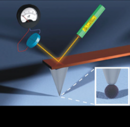
Schematic overview of the atomic force microscope. An enlargement of the tip of the needle that scans the virus is shown at the bottom right.
More information: The article Mechanics of Bacteriophage maturation is the 30th of January 2012 in Proceedings of the National Academy of Sciences.
Provided by University of Amsterdam


