October 20, 2020 feature
Microscopy with undetected photons in the mid-infrared region

Thamarasee Jeewandara
contributing writer

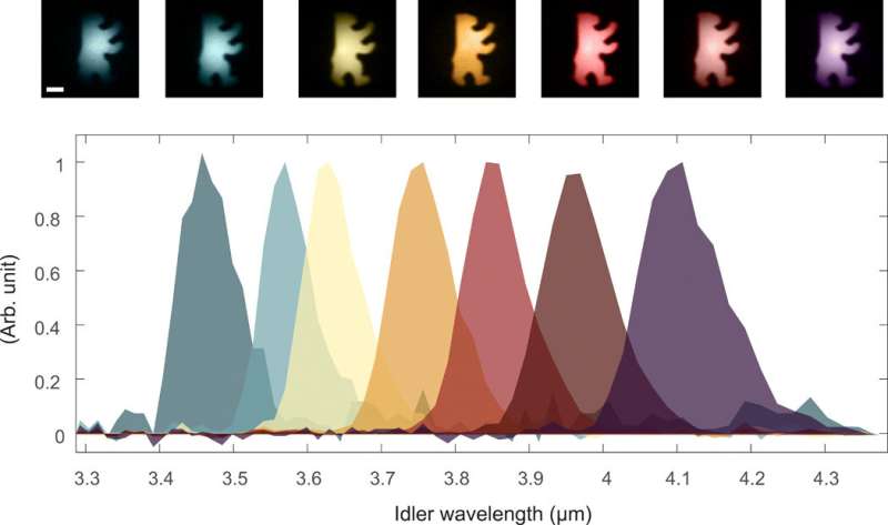
Microscopy techniques that incorporate mid-infrared (IR) illumination holds tremendous promise across a range of biomedical and industrial applications due to its unique biochemical specificity. However, the method is primarily limited by the detection range, where existing mid-infrared (mid-IR) detection techniques often combine inferior methods that are also costly. In a new report now published on Science Advances, Inna Kviatkovsky and a research team in physics, experimental and clinical research, and molecular medicine in Germany, found that nonlinear interferometry with entangled light provided a powerful tool for mid-IR microscopy. The experimental setup only required near-IR detection with a silicon-based camera. They developed a proof-of-principle experiment to show wide-field imaging across a broad wavelength range covering 3.4 to 4.3 micrometers (µm). The technique is suited to acquire microscopic images of biological tissue samples at the mid-IR. This work forms an original approach with potential relevance for quantum imaging in life sciences.
Mid-IR imaging
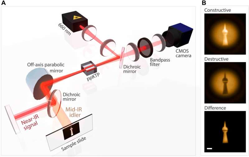
Microscopy and mid-IR imaging have broad ranging applications across , and . For example, researchers can use mid-IR light to sense the distinct rotational and vibrational modes of specific molecules as a "spectral fingerprint," to overcome the need for labeling. Such label-free and non-invasive techniques are important for bioimaging procedures in largely unaltered living tissues. Fourier transform IR spectroscopic imaging is a that heavily depends on broadband IR sources and detectors. The IR detectors are, however, technically challenging, costly and sometimes require cryogenic cooling. To bypass the need for IR detectors, researchers must develop coherent Raman and methods. In a markedly different approach, they used the with widely different wavelengths that do not require laser sources or detectors at the imaging wavelength. In this work, Kviatkovsky et al. used highly multimodal as a powerful tool for microscopic imaging in the mid-infrared region with only a medium powered visible laser and standard custom metallic-oxide semiconductor (CMOS) camera. They derived explicit formulas for the field-of-view and resolution of wide-field imaging with highly nondegenerate photon pairs.

The experimental setup
The scientists developed a nonlinear interferometer by double passing a periodically poled (ppKTP) crystal in a folded (an interference pattern). The pump passed the crystal twice to generate a single pair of signal and idler photons through (SPDC) - a non-linear optical process where a photon spontaneously splits into two other photons of lower energies in an optics lab. The SPDC method forms the foundation of many quantum optical experiments in labs at present, ranging across quantum cryptography, quantum metrology to even facilitate the testing of fundamental laws of quantum mechanics. The signal and idler modes aligned after the first pass of the crystal to propagate back for the second pass and perfectly overlap to generate biphotons. Kviatkovsky et al. measured the interference by looking at the signal photons with a CMOS camera, without including complex or cost-intensive components to realize such a setup. The team engineered the nonlinear crystal for highly nondegenerate signal and idler wavelengths and selected the idler wavelengths using . In this way, the experiment allowed simultaneous retrieval of the spatially resolved phase and amplitude information of a sample and the team characterized the mid-IR imaging properties with an off-the-shelf CMOS camera to detect and acquire microscopic images of a biological sample.

Experimental characterization and proof-of-concept
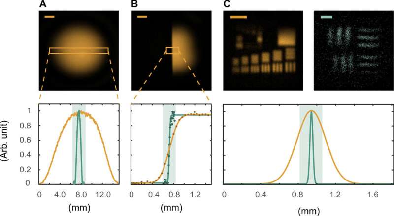
During the initial characterization of the imaging technique, Kviatkovsky et al. placed both mirrors of the interferometer at the far-field of the crystal and then placed the sample to be imaged on the idler mirror. The unmagnified configuration provided a straightforward process to characterize the imaging capacity of the system, although with limited resolution. The scientists illuminated a U.S. Air Force (USAF) clear path resolution target, where the resulting values were consistent with a theoretical framework generalized from . They combined the highly broadband nature of the down-conversion source with tight energy correlations shared between the signal and idler to easily allow hyperspectral imaging. During proof-of-concept demonstrations, they used a tunable interference filter with a 3.5 nm bandwidth immediately before detection and achieved enhanced spectral resolution with narrower filtering.
Using the method for bioimaging
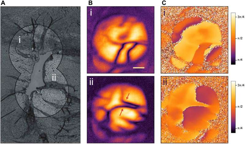
The team showed the potential of the method to investigate biological samples by using an unstained sample of a mouse heart. They obtained mid-IR images by axially scanning the interferometer displacement inside the coherence length and extracted the visibility and phase of the interference signal for each pixel. The results eliminated any ambiguity between loss and destructive interference that could arise in a single-shot measurement. The work permitted straightforward reconstruction of the wide-field phase-contrast images. The resulting images showed a portion of the , the inner-most layer lining the heart ventricles in dark purple to indicate high photon absorption. The layer separated the ventricle and the ; the cardiac muscle that consists the bulk of the heart tissue. The clarity of imaging highlighted the high tolerance of the presented imaging method to overcome loss and scattering.

Real-world promise
In this way, Inna Kviatkovsky, and colleagues showed how mid-IR imaging with nonlinear interferometry played a significant role in real-world imaging tasks that require cost-efficient components for frugal science. The team achieved an imaging feature down to the scale of 35 microns, where extended hyperspectral imaging was uncomplicated due to the use of a broadband (SPDC) strategy. The team showed real-world promise of this new method through non-destructive biological sensing while imaging a wet biological sample with low sample illumination. The strategy allowed any information carried by an idler photon to be perfectly transferred to the signal photon. Although the spatial resolution of this work was still higher than that anticipated for state-of-the-art mid-IR systems, extensions to accomplish increased imaging capabilities were straightforward.
The team showed nonlinear interferometry with experimentally entangled photons to provide a powerful and cost-effective method for microscopy in the mid-IR region. The work harnessed the maturity of silicon-based near-IR detection technology for mid-IR imaging with exceptionally low-light level illumination. The work can be extended to hyperspectral imaging across the microscale. As proof of concept, the scientists imaged a biological sample using quantum light to reveal morphological features with high resolution. The results will pave way for broadband, hyperspectral mid-IR spectroscopy with wide-field imaging for varying applications in biology and biomedical engineering.
Written for you by our author —this article is the result of careful human work. We rely on readers like you to keep independent science journalism alive. If this reporting matters to you, please consider a (especially monthly). You'll get an ad-free account as a thank-you.
More information: Kviatkovsky I. et al. Microscopy with undetected photons in the mid-infrared , Science Advances,
J.-X. Cheng et al. Vibrational spectroscopic imaging of living systems: An emerging platform for biology and medicine, Science (2015).
Gabriela Barreto Lemos et al. Quantum imaging with undetected photons, Nature (2014). 0
Journal information: Science Advances , Science , Nature
© 2020 Science X Network


